



Overview
The retina is like a camera film: it functions by capturing the light from objects. The image directed onto the retina is then transmitted to the brain where it is processed. Structurally speaking, the retina is more like a part of the brain with no capacity for self-repair or transplantation. In the centre of the retina is the macula, which is responsible for central and colour vision. A healthy macula allows us to see objects in fine detail, read and see colours. If left undiagnosed and untreated, all types of maculopathies can lead to severe consequences. Thanks to advances in clinical technology and a better understanding of macular diseases, those that were considered incurable in the past can now be treated by surgery.
Risk Factors of Macular Diseases
- Genetics – with family history
- High myopia (above 600 degrees)
- Diabetes mellitus
- Hypertension
- Hyperlipidemia
- Smoking
- Cardiovascular disease
- Aging
Symptoms and Diagnosis
Symptoms of Macular Diseases
- Absence of symptoms, especially when they affect one eye only or are in slow progression
- Blurred central vision
- Central visual field defects
- Distorted images
Diagnostic Tests
If you experience any symptoms, it is important that you seek medical attention as soon as possible. Testing may include
- Dilated eye examination by ophthalmologist
- Optical coherence tomography (OCT)
- B scan
- Fluorescein fundus angiography (FFA)
- Indocyanine green angiography (ICG)
Procedures and Treatments
Latest Developments in Maculopathy Treatment
Treatment of Macular Hole
A macular hole is a break in optic nerve tissues in the centre of macula. It is mostly age-related or caused by severe myopia. The standard treatments are subtotal pars plana vitrectomy with removal of the posterior hyaloid, followed by removing the internal limiting membrane (Photo 1) and gaseous and fluid exchange. Most patients are not required to maintain a face down posture post-operatively. The success rate is over 90%.
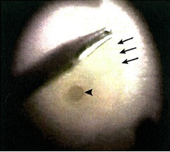
Photo 1: During surgery (the right eye of the patient from the surgeon's perspective): the internal limiting membrane near the macular hole (arrow) is lifted (arrow) using an intraocular forceps.
Treatment of Proliferation of Macular Epiretinal Membrane
Proliferation of macular epiretinal membrane (Photo 2) is one of the common eye diseases in Hong Kong, the treatment of which is to remove the vitreous body and epiretinal membrane. Outcomes can be improved with internal limiting membrane removal. The recurrence rate after first surgery is low.
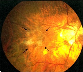
Photo 2: Macular epiretinal membrane disease in a patient with severe myopia (before surgery).
Treatment of Maculoschisis
Those with myopia of over 600 degrees have severely elongated eyeballs, which stretch the macula thin. The macular split could be aggravated due to further stretching by abnormal vitreous body and epiretinal membrane, leading to loss of vision. The split might deteriorate into macular holes (Photo 3A) and even macular retinal detachment if left untreated. With Optical Coherence Tomography (OCT), it can now be detected and diagnosed in its early stage with high accuracy (Photo 3B). The treatment is pars plana vitrectomy with removal of the posterior hyaloid, epiretinal membrane and internal limiting membrane followed by gas injection. They can resolve maculoschisis in most patients. Most patients are not required to maintain a face down posture post-operatively.
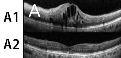
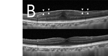
Photo 3: Optical Coherence Tomography (OCT):
(A1) Extensive macular split, macular detachment and loss of macular depressions are detected under OCT before surgery.
(A2) Macular split improves significantly 6 months after surgery.
(B) Early stage of maculoschisis is detected under OCT.
Treatment of Neovascular Membranes Caused by Age-Related and Severe Myopia Macular Degeneration
Photodynamic therapy was once the standard treatment of neovascular membranes caused by age-related and severe myopia macular degeneration. It has a limited effect of maintaining the remaining vision or deterring vision loss. Currently, intraocular injection (incl. Lucentis, Avastin, Eylea, Beovu, Vabysmo and steroids) is the common practice. It can restore vision to a certain extent in most patients. The procedure is safe, and side effects are rare.
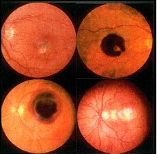
Photo 4: Age-related macular degeneration - bleeding
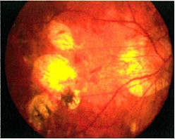
Photo 5: Severe myopia macular degeneration
Contact Us

Happy Valley
- Address:
- 4/F, Li Shu Fan Block,
Hong Kong Sanatorium & Hospital,
2 Village Road, Happy Valley, Hong Kong
- Service hours:
-
Monday to Friday: 9:00 am - 5:00 pm
Saturday: 9:00 am - 1:00 pm
Closed on Sundays and Public Holidays
Consultation by Appointment
- Tel.:
- (852) 2835 8880
- Email:
- [javascript protected email address]
- Fax:
- (852) 2892 7510
- WhatsApp:
-
(852) 2835 8890
(For non-emergency cases)

Admiralty
- Address:
- 21/F, One Pacific Place,
88 Queensway, Hong Kong
- Service hours:
-
Monday to Friday: 9:00 am - 5:00 pm
Saturday: 9:00 am - 1:00 pm
Closed on Sundays and Public Holidays
Consultation by Appointment
- Tel.:
- (852) 2855 6700
- Email:
- [javascript protected email address]
- Fax:
- (852) 2523 7660
- WhatsApp:
-
(852) 2855 6700
(For non-emergency cases)

Admiralty
- Address:
- 21/F, One Pacific Place,
88 Queensway, Hong Kong
- Service hours:
-
Monday to Friday: 9:00 am - 5:00 pm
Saturday: 9:00 am - 1:00 pm
Closed on Sundays and Public Holidays
Consultation by Appointment
- Tel.:
- (852) 2855 6699
- Email:
- [javascript protected email address]
- Fax:
- (852) 2523 7660
- WhatsApp:
-
(852) 2855 6699
(For non-emergency cases)

Happy Valley
- Address:
- 6/F, Central Block,
Hong Kong Sanatorium & Hospital,
2 Village Road, Happy Valley, Hong Kong
- Service hours:
-
Consultation by Appointment
- Tel.:
- (852) 2835 8890
- Email:
- [javascript protected email address]
- Fax:
- (852) 2892 7538

Admiralty
- Address:
- 22/F, One Pacific Place,
88 Queensway Hong Kong
- Service hours:
-
Consultation by Appointment
- Tel.:
- (852) 2855 6700
- Email:
- [javascript protected email address]
- Fax:
- (852) 2892 7429